
Kidney Stone Treatment in Delhi, India
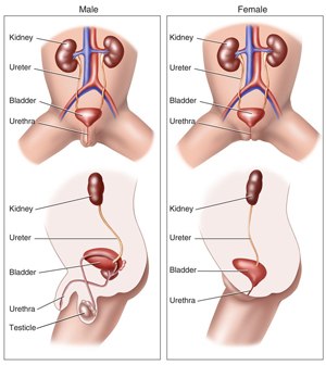
The urinary tract consists of a pair of kidneys which are situated close to the back of a person. The primary function of the kidney is to filter blood and form urine which contains various waste products of the body. The urine which forms in the kidney briefly stays in a part of the kidney which is called as a pelvis. From the renal pelvis, the urine is transported to the urinary bladder via a thin muscular tube which is called a ureter. The urine is stored in the urinary bladder and from here it is periodically discharged to the exterior through a tube which is called the urethra. In males, the urethra passes through the penis and thus it is much longer than in females. The prostate is an internal organ which is peculiar to males and surrounds the urethra at the base of the bladder. In the younger age, it contributes to the seminal fluid but as the male ages, it increases in size and creates an obstruction to the flow of urine. It can also become cancerous.
Kidney Stone Surgery India
An overview
The urine which is filtered from the kidneys contains minerals like calcium, phosphorus, sodium, uric acid, oxalates.
If the saturation of these minerals increases in the urine or if the flow of urine is retarded there is a risk of deposition and aggregation of these minerals resulting in stone formation.
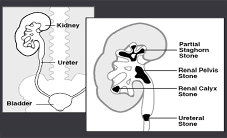
The fate of these stones is unpredictable. They can remain silent/slowly enlarge and cause damage to the kidney. If they move into the ureter or get stuck in any part of the urinary system they cause severe pain which is due to increased pressure on the closed urinary system. Stones less than 4 mm usually pass spontaneously.
Symptoms of urinary calculi
Pain: it characteristically starts from the back or lower part of the abdomen and spreads to the entire side. Sometimes it is also referred to the external genitalia and thighs The location and severity of the pain depend upon the location of the stone in the urinary tract, the degree of obstruction, and kidney function. Poorly functioning kidneys cause less pain.
Other symptoms include
- Blood in urine
- Fever which indicates infection
- Urinary trouble
- Nausea and vomiting
- Tenderness in the abdomen and on the sides of the spine
Diagnosis
Ultrasound: A widely available investigative modality, detects stones in the kidney with reasonable accuracy. It Can also detect the dilated kidney and ureter thus indicating an obstruction in the urinary tract. It is less sensitive in localizing ureteral stones.
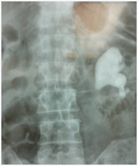
X-ray KUB: It can detect stones that are radio-opaque (i.e. Seen on x-ray). However, the accuracy of x-ray depends upon the number of gases present inside the abdomen and density of the stone.
Intra Venous pyelography(IVP): This test involves taking a series of x-rays after injecting contrast (dye) into the vein. The contrast flows through the veins and is excreted by the kidneys. As it can be seen on the x-ray, the whole urinary tract containing the contrast can be delineated. Any obstruction in the urinary tract can be picked up. It can also indicate the kidney function and level of stone. A normal kidney function test is a prerequisite to performing IVP.
CT Scan: A non-contrast CT scan is the fastest and most accurate investigation for urinary calculi. It can be performed without the need for contrast and thus is safe in patients with a history of radio-contrast allergy. It also gives an opportunity to screen other abdominal organs at the same time.
Treatment of kidney stones
- Size of stone
- Location
- Kidney function
- Presence of infection
- Anatomy of the urinary tract
Treatment modalities
- ESWL or Extra-corporeal Shock Wave Lithotripters
- PCNL or Per Cutaneous Nephrolithotomy
- URS or Ureterorenoscopy
- RIRS or Retrograde Intrarenal Surgery
- Laparoscopic surgery
- Open surgery
ESWL
This procedure focused shockwaves from outside the body to crush stones in the urinary tract. The stone breaks according to its hardness. Soft stones break into sand-like particles which pass in the patient’s urine. It is used for stones up to 1 cm in the kidney and upper parts of the ureter. More than one session may be required for breaking the stone. It should not be used in pregnant women. Sometimes the stone pieces get stuck while coming out and may require a ureteroscopic removal or temporary insertion of a stent.
PCNL
This is one of the most common methods currently employed in the removal of kidney stones. This procedure involves establishing a track from the skin into the kidney through which instruments are passed inside and the stone is broken into smaller pieces and removed. It can be used for stones of any size. Multiple tracts may be required for complex stones. The incision used for making these tracts is 5 to 10 mm. and does not require to be stitched usually. Hospitalization is usually required for 2 to 3 days and the patient can resume normal work in a weeks time. The only major risk of this operation is bleeding which is unpredictable and can occur in up to 1% of the patients requiring a blood transfusion.
URS
As the name suggests it involves insertion of the fine instrument through the penis into the ureter. The stone can be visualized, broken by the holmium laser and removed.
It is indicated for the treatment of ureteric stones which are accessible to the ureteroscope. Sometimes in the presence of infection or very tight ureter, the procedure is done in two stages. A ureteric stent is usually required to be placed at the end of the procedure which is removed after two weeks.
RIRS
This is also a form of ureteroscopy but here the instrument used is flexible and thus can negotiate most parts of the ureter and can reach into the kidney from below.
It obviates the need for puncturing the kidney for small renal stones which cannot be taken care of by simple lithotripsy.
Open/Laparoscopic Surgery
These modalities are rarely used currently, however they have their own place in certain special situations.
Prevention of stone disease
No discussion on urinary stones is complete without information on their prevention. The recurrence rates are very low in the first year but over a period of ten years 30 to 40%, people have recurrences.
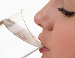
General preventive measures are increased intake of fluids, citrus fruit juices and limiting intake of salt, meat, chocolate, dry fruits, etc. Urinary tract infections and other abnormalities should also be corrected.
The patient should drink enough fluids so as to produce a urine output of more than 2 liters.
Special tests like stone analysis and metabolic workup help in finding the cause of kidney stones in an individual.
Contrary to the popular belief beer is not helpful in the prevention and expulsion of stones.
Services